Research Highlights
Quantum Tomography of a Molecular Bond in Ice
I. U. Goldschleger, M. van Staveren, V. A. Apkarian, J. Chem. Phys. 139, 034201 (2013); DOI: 10.1063/1.4813437
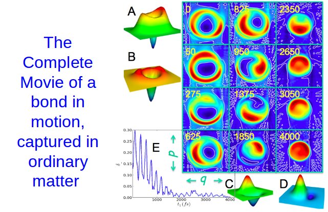 |
Selected frames of the moving bond captured in phase space, at 5 fs per frame. The abscissa (q) is the bond length of Bromine (0.1 Å full scale), and ordinate (p) is the momentum of the moving bond. The images are the tomographically reconstructed Wigner distribution function (WDF), which provides the complete uncertainty limited quantum mechanical description of a dynamical system. The white line is the zero-contour, which shows the strictly quantum effect of a negative volume in the WDF, The graph is the time dependent zero-volume, which decays as the system relaxes to v = 0. (The complete video will appear on line, linked through the manuscript.)
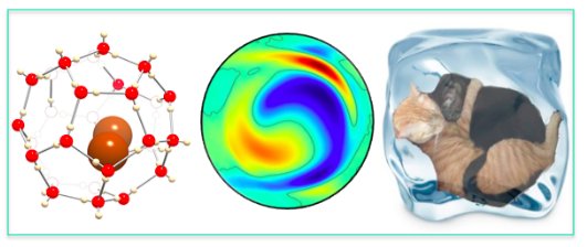 |
The measurements are carried out in a sliver of dirty ice: Bromine doped ice, at T = 120 K. The molecule creates a cage around itself (clathrate) as show in the first image. Despite the environment, the molecular motion is fully quantum mechanical, which is certified by the negative hole in the WDF and the observed t-dependent wavefunctions (middle). The negative hole arises only from quantum interference, and such states are referred to as “cat” states.
Orbiting Orbital: Visualization of Vibronic Motion at a Conical intersection Or the Molecular Hoola Hoop
J. Lee, S. M. Perdue, A. Rodriguez-Perez, P. Z. El-Khoury, K. Honkala, V. A. Apkarian, J. Phys. Chem. 2013. DOI: 10.1021/jp311894n
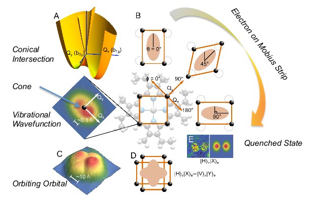 |
Images of motion of electron and vibrational deformation, and the first visualization of a conical intersection, by imaging the unpaired e- of Zn(II)-etioporphyrin radical anion, which is Jahn-Teller active. Its degenerate orbital is dragged into orbit by pseudo-rotation of the molecular skeleton. in orbit, it looks like a clover. When the motion is quenched by applying bias, the e- looks like a dumbbell. The 0.1 Å vibration of the skeleton sets the e- on 15 Å hoola hooping orbit.
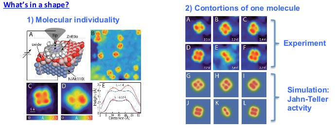 |
Shapes and reactivities of molecules are determined by their frontier orbitals. Under the pristine conditions of cryo-UHV, on an inert film of Al2O3, the individuality of molecules is apparent by the variation in their shapes (1). The same molecule undergoes contortions upon polarizing the substrate (2). The effect is dramatic in the radical anion, but fully reproduced as Jahn‐Teller activity.
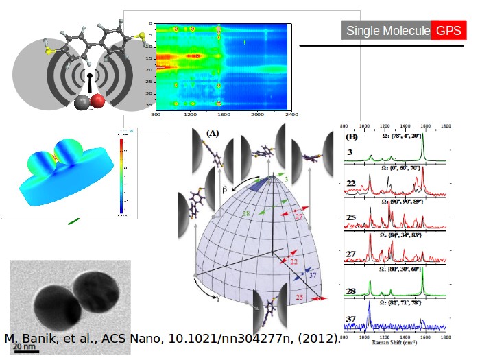
A molecular GPS, AC/DC voltmeter, and a rectifying antenna consisting of a chemically engineered nano-dumbbell
M. Banik, P. Z. El-Khoury, A. Nag, A. Rodriguez-Perez, N. Guarrottxena, G. C. Bazan, and V. A. Apkarian
Project Outcome: This research demonstrates a “global positioning system” for tracking single molecules in 3D space by equipping them with nano-dumbbell antennae; and the use of Raman spectra as a voltmeter to characterize AC and DC fields on nanometric scale. When a small asymmetric molecule is trapped in the junction of metallic nano-spheres, the dumbbell acts as a rectifying antenna (rectenna).
Impact & Benefits: The ability to remotely track single molecules in 3D space is an essential step toward chemistry of individual molecules. Rectennas fulfill ideal design requirements of a photocell, with enhanced spectral response and conversion efficiency over existing cells. The demonstrated principles should advance the state-of-the-art in harnessing the energy of the sun.
Background & Explanations: In an approach analogous to GPS, by equipping molecules with nano-antennae and taking advantage of the tensor nature of Raman scattering, it is demonstrated that fluctuations in surface enhanced Raman spectra (SERS) of single molecules can be used to locate them in 3D space, as a function of time. Relying on the vibrational Stark shift of CO, it’s SERS spectrum serves as a molecular voltmeter, which is used to characterize the plasmonic fields at the junction of silver nano-spheres. The observed DC field of 1 V/Å, characterizes the dumbbell as a rectenna with a spectral response that covers the visible to the IR, as a function of junction gap.
Publications Relevant to Project Outcome: " Surface-Enhanced Raman Trajectories on a Nano-Dumbbell: Transition from Field to Charge Transfer Plasmons as the Spheres Fuse" ACS Nano, DOI:10.1021/nn304277n, (2012).
  |
Nanocircuits Measure Bacteria-chomping Lysozyme Action
Y. Choi, I. S. Moody, P. C. Sims, S. R. Hunt, B. L. Corso, I. Perez, D. Seitz, L. Blaszcazk, G. A. Weiss, P. G. Collins
Project Outcome:
This research proves the feasibility of recording single molecule chemistry using ultrasmall electronic devices. The project built devices out of single lysozyme molecules, and then watched the electrical signal that resulted as the enzyme went about its normal activity of chopping apart bacteria.
Impact & Benefits:
The diagnosis of diseases or the discovery of new medicines both require that scientists be able to unravel chemical reactions happening between complex molecules. This research has invented a new diagnostic tool that allows reactions to be recorded and analyzed with molecule-by-molecule precision.
Background & Explanations:
By harnessing the chomping ability of lysozyme and latching it to a carbon nanotube, researchers at UCI created the most precise chemical-reaction monitor ever developed. Lysozyme is a bacteria-munching enzyme found in humans, and when it encounters particular bacteria it rips into them with a rapid chomping motion. The research here proved that a nanotube sensor could convert this chomping action into an electric signal, making it possible to record each biochemical step made by the enzyme. Besides shedding new light on the activity of this particular molecule, the research proves the possibility of recording chemistry molecule by molecule. This invention can be put to use as part of a toolbox for understanding complex molecules and their chemistry. For example, it enables researchers to quantify the effects of mutations, observe interference by pharmaceuticals, and measure the binding of molecules at docking sites. Future applications may include sensors, detector technologies, and diagnostic tools for health care.
Publications Relevant to Project Outcome:
"Single-molecule lysozyme dynamics monitored by an electronic circuit" Y. Choi, I. S. Moody, P. C. Sims, S. R. Hunt, B. L. Corso, I. Perez, G. A. Weiss, P. G. Collins, Science, 335, 319 (2012).
"Single-molecule dynamics of lysozyme processing distinguishes linear and cross-linked peptidoglycan substrates" Y. Choi, I. S. Moody, P. C. Sims, S. R. Hunt, B. L. Corso, D. Seitz, L. Blaszcazk, P. G. Collins, G. A. Weiss, J. Am. Chem. Soc., 134, 4 (2012).
> View Article From Science Volume 335
> View Article From The Journal of the American Chemical Society
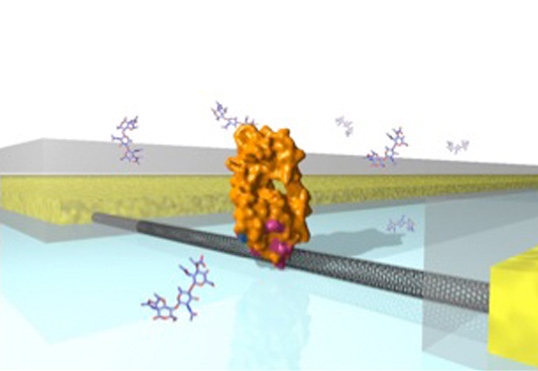
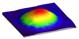
A lysozyme nanocircuit. |
Spatially and Spectroscopically Resolved Molecules on a Surface
N. Jiang, E. Foley, J. Klingsporn, M. Sonntag, N. Valley, J. Dieringer, T. Seideman, G. C. Schatz, M. C. Hersam, and R. P. Van Duyne
Project Outcome:
Professor Van Duyne’s group has combined scanning tunneling microscopy (STM) with tip-enhanced Raman spectroscopy (TERS) to both spatial image at sub-nm resolution and resolve chemical bonding information of large polyatomic molecular adsorbates on a metal surface. Theoretical electronic structure calculations and simulated Raman spectra were carried out and showed strong agreement between theory and experiment of the vibrational modes of the absorbate.
Impact & Benefits:
The ability to resolve multiple vibrational peaks while simultaneously imaging at the sub-nm molecular levels allows the chemical relationship between polyatomic adsorbates and solid surfaces to be quantitatively interrogated. This technique presents new opportunities for the study of catalysis and other molecule-surface related phenomena necessary for the understanding of important innovations. For example, the adsorbate studied, copper phthalocyanine (CuPc), has potential applications in organic light-emitting diodes, thin-film transistors, as well as other optical and electronic devices.
Background & Explanations:
Tip-enhanced Raman spectroscopy (TERS) combined with surface imaging techniques, such as STM, can provide quantitative information on surface morphology, binding sites, and the vibrational fingerprint of adsorbates on a solid surface. The Van Duyne’s group experimental setup allows for unprecedented spatial and spectroscopic resolutions, with the full potential of this technique not yet realized.
Publications Relevant to Project Outcome:
"Observation of Multiple Vibrational Modes in Ultrahigh Vacuum Tip-Enhanced Raman Spectroscopy Combined with Molecular-Resolution Scanning Tunneling Microscopy" N. Jiang, E. Foley, J. Klingsporn, M. Sonntag, N. Valley, J. Dieringer, T. Seideman, G. C. Schatz, M. C. Hersam, and R. P. Van Duyne, Nano Lett., ASAP, DOI: 10.1021/nl2039925. (2012).
> View Article From American Chemical Society
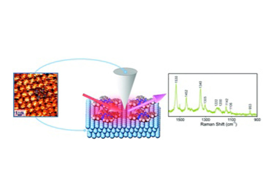
Spatially and Spectroscopically Resolved Molecules on a Surface” Caption: Schematic |
We illustrate the electric field lines (red/yellow) and |
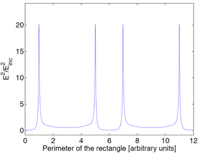
We show the square of the electric field, normalized to |
Rapid Vibrational Imaging With Sum Frequency Generation Microscopy
Varun Raghunathan, Yang Han, Olaf Korth, Nien-Hui Ge and Eric O. Potma
October 2011
Project Outcome:
The Ge and Potma groups joined forces in constructing a new type of vibrationally resonant sum frequency generation (VR-SFG) microscope. VR-SFG microscopy enables a detailed look at biologically relevant samples and molecules at interfaces based on vibrational sensitivity. The CaSTL researchers have successfully shown sub-micrometer resolution directly correlating orientation, morphology, and spectroscopic properties of collagen fibers.
Impact & Benefits:
The Potma and Ge groups have overcome many of the existing shortcomings of the VR-SFG imaging technique by using a high-repetition-rate pico-second light source and a collinear excitation geometry. This high-speed laser scanning microscope set-up has the potential to reveal precise spectroscopic information about molecules at interfaces, such as bond-specific information, with sub-micrometer spatial resolution.
Relevant Publications:
"Rapid vibrational imaging with sum frequency generation microscopy" V. Raghunathan, Y. Han, O. Korth, N.-H. Ge and E. O. Potma. Optics Letters 36 (19), 3891-3893 (2011).
> View The Article From Optical Society of America
Resolving Individual Molecules Using Surface-Enhanced Femtosecond Stimulated Raman Spectroscopy
Renee R. Frontiera, Anne-Isabelle Henry, Natalie L. Gruenke and Richard Van Duyne
March 2011
Project Outcome:
Professor Van Duyne's group, in collaboration with coworkers at UC Irvine, has succeeded in integrating extremely fast time-resolved spectroscopy to dynamically probe nanomaterials. Using the novel technique of Surface-Enhanced Femtosecond Stimulated Raman Spectroscopy (SE-FSRS), we are able to detect extremely small quantities of molecules bound to nanoparticles. In the bottom right image, we show the first ever SE-FSRS spectra; these spectra of bipyridine molecules bound to gold nanoparticles are highly resolved. We are thus able to examine molecules on the billionth of a meter length scale with time resolution of up to a million billionth of a second, achieving the ability to examine chemistry at the limits of space and time.
Impact & Benefits:
This research is aimed at understanding the molecular processes that occur in novel nanomaterials that are becoming important players in solving the global energy crisis. New solar cell technologies that use these novel materials, such as dye-sensitized solar cells, are very promising due to the use of inexpensive materials and solution-based processing. By examining the motions of small number of molecules after they absorb light and convert it to energy, we can determine the optimal chemistry and molecular orientations that are needed to achieve a high-energy conversion efficiency. This fundamental understanding can be used to rationally design new materials, molecules, and solar cell device architectures.
Relevant Publications:
"Surface Enhanced-Femtosecond Stimulated Raman Spectroscopy" R. R. Frontiera, A.-I. Henry, N. L. Gruenke, and R. P. Van Duyne Journal of Phys. Chem. Lett. 2 (10), 1199-1203 (2011)
> View Article From The Journal of Physical Chemistry Letter

Experimental setup showing methods for creating |
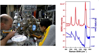
Left: CaSTL researchers developing new techniques for |
Lighting Up the Interior of a Single Molecule
Chi Chen, Ping Chu, Christian Bobisch, Doug L. Mills, and Wilson Ho
January 2011
Project Outcome:
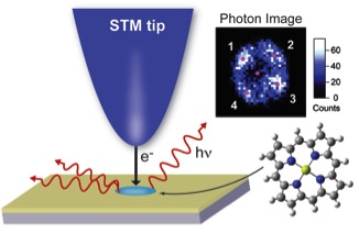
Schematic of imaging photon transitions of a single magnesium porphine molecule at a submolecular resolution using a Scanning Tunneling Microscope (STM). Electrons that tunnel from the STM tip to the molecule, excite the molecule that then relaxes via a radiative transition (fluorescence) that can be detected. The resulting Photon Image (inset) is spatially resolved by scanning the tip and reveals that the optical transition occurs at the outer porphyrin rings.
In the past 20 years, optical experiments have reached single molecule sensitivity and removed ensemble averaging associated with variations in the molecular environment. However, each emitting molecule appears as a beacon of light without internal structure due to the limited spatial resolution. The Ho group has successfully resolved intramolecular optical transitions in real space at submolecular resolution. By tunneling electrons from a tip of a scanning tunneling microscope (STM) to an individual magnesium porphine molecule, they were able to excite the molecule and image the corresponding fluorescence with sub-Ångstrom precision. The molecule was mapped by collecting light for different tip locations over the molecule. Single-point spectroscopy results show that the optical transition occurs at the outer porphyrin rings of the molecule, and that the central magnesium ion is not involved in the luminescence process. The images also reveal a twofold symmetry for the four porphyrin rings when light is collected from individual vibronic states of two molecular orbitals that are spatially orthogonal to each other. Besides the optical emission mapping, the Ho group also used the STM to determine electronic state information about the molecule. Combining these experimental results with theoretical calculations by D. L. Mills' group, the fundamental aspects of molecular radiative transitions are explained.
Impact & Benefits:
This technique combines topographic imaging, electronic and optical spectroscopy, and photon mapping to greatly advance the field of single molecule spectroscopy and microscopy. This is the first instance of spatially resolving photon imaging of the vibronic emission of a single molecule. With this approach, intrinsic emission properties of a single molecule can be visualized, and the effects of its local environment can be accounted for. This method leads to unprecedented opportunities to study fundamental processes that take place below the nanometer scale, providing a new window to observe the coupling of electron and light in a molecule that forms the basis for dye sensitized solar energy conversion, organic light emission, and photocatalytic chemistry.
Relevant Publications:
Chi Chen, Ping Chu, C. A. Bobisch, D. L. Mills, and W. Ho. "Viewing the Interior of a Single Molecule: Vibronically Resolved Photon Imaging at Submolecular Resolution" Physical Review Letters 105 (2010): 217402.
Highlighed By:
APS Viewpoint: Marina Pivetta. "Mapping the luminescence of a single molecule." American Physical Society – Viewpoint, Physics 3 (2010): 97.
Nature Research Highlights: "Optical Physics: A peek at a molecule’s guts." Nature 468 (2010): 602-603.
Optics and Photonics News: Yvonne Carts-Powell "Lighting Up the Inside of a Single Molecule." Feb. 2011
Optics and Phontonics Focus: "The Ultimate Resolution." Feb. 2011
> View Article From The American Physical Society
Lightning Without Thunder
Joonhee Lee, Shawn M. Perdue, Desiré Whitmore, and V. Ara Apkarian
November 2010
Project Outcome:
The Apkarian group has demonstrated unprecedented thermal management of an STM tip under irradiation with femtosecond laser pulses, keeping its expansion stable to less that 1/100 of an atom, while modulating the output current. They achieved this through a novel "double beat" method that maintains a constant heat load on the tip, yet still gives a signal modulation for increased sensitivity. In this method, a laser pulse is split in two, with one copy being frequency shifted from the other by an acousto-optic modulator (AOM). When the two pulses are recombined in an interferometer, they almost cancel out, but leave that frequency shift as the first beat. This new frequency then beats with the repetition rate of the laser for the second beat. The use of a double beat brings the signal modulation into the base-band, where it can be amplified by a high gain preamplifier.
The key to eliminating the thermal shaking, or thunder, is in cross-polarizing the two pulses. In this way, there is no interference between them until they impinge on the STM tip itself. The tip then sees the pulse trains individually, which come too fast for it to respond thermally. However, the tunneling electrons, or lightning, do see the interference in the electric field, and are thus modulated themselves. Lightning without thunder!
Impact & Benefits:
The double beat method opens the possibility of ultrafast triggering of STM, and consequently, videography of single chemical events. Although this is the primary goal of this research group, the applications of this new method extend much further. Light plays a key role in many chemical processes, and the ability to optically stimulate a sample while imaging it on an atomic level enables access to a new level of the experimental study of single molecules.
Background & Explanations:
To explore chemistry at the measurable limits of space and time, Ångstroms and femtoseconds, the major challenge has been in combining ultrashort laser pulses with scanning tunneling microscopy (STM). Specifically, the thermal modulation of the gap between the STM tip and the sample has been difficult to control. To be useful in microscopy, the gap must be stable to within 0.01 Å. That's more than 30 times smaller than a hydrogen atom! Even a 1 mW laser, like a laser pointer, will cause a typical tip to expand by 10,000 times this limit. The double beat method simultaneously enables gap stability and signal modulation.
Relevant Publications:
Lee, Joonhee, Shawn M. Perdue, Desiré Whitmore, and V. Ara Apkarian. "Laser-induced scanning tunneling microscopy: Linear excitation of the junction plasmon." Journal of Chemical Physics 133 (2010): 104706.
Highlighted By:
Virtual Journal of Nanoscale Science & Technology, September 20, 2010
Virtual Journal of Ultrafast Science, October 2010
> View Article From The Journal Chemical Physics

A scanning tunneling microscope (STM) tip above a silver |
STM image of a single silver atom on a NiAl surface. |
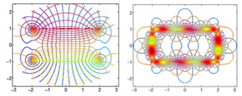
Plasmon Resonances of Nanowires and Their Consequences
Alejandro Jara, Rodrigo Arias and Doug L. Mills
February 2010
Project Outcome:
A major challenge in the study of single molecules and very small entities by laser spectroscopy is to find the means of boosting the signal to detectable levels. This may be achieved though exploitation of resonance modes of nanoscale metallic entities referred to as plasmons. When such a resonance is excited by a laser beam, the electric fields in the near vicinity of the nanostructure which supports the plasmon can be enhanced greatly over that present in the beam itself, thus greatly boosting the strength of any non linear optical process of interest. A plasmon is a collective, coherent resonant response of the conducting electrons in the nanoscale structure, which is illuminated on resonance to provide the field boost.
It is imperative to have the means to explore which geometries are maximal for providing the field enhancement just described. Here theory provides crucial input. In this project, we have formulated a new and novel theoretical approach that allows us to explore the nature of the plasmon normal modes of nanowires of arbitrary cross section. We are able to map out the electric field lines of each individual plasmon modes, as illustrated in Image 1. Our formalism is unique in this regard; this cannot be done, for instance, with the standard FDTD or COMSOL software used by many researchers. Our formalism also enables us to calculate, for any real nanowire, its response to an external laser field and explore in a fully quantitative manner the nature of the electric fields, which surround the object.
The information in Image 1 and Image 2 above is directed toward the nanowires of rectangular cross section studied in the lab of Prof. Eric Potma and Prof. Reg Penner. We see that for nanowires of this cross section, the strong field enhancement occurs very near the corners (Image 2) when the fundamental plasmonmode is excited. The origin of this effect is in polarization charges located at the corners of the nanowire, illustrated for example in Image 1.
Impact & Benefits:
Our new formalism, already applied to a geometry of direct interest to an experimental phase of the CaSTL program, can be readily employed by research groups elsewhere to explore the plasmonic response of nanowires of diverse cross section. Thus, it will enable other groups to optimize the strength and nature of laser fields near diverse nanostructures. In our own program, we have initiated the study of nanocrescents and related structures.
Relevant Publication:
"Plasmons and The Electromagnetic Response of Nanowires", Alejandro Jara, Rodrigo Arias and D. L. Mills, Physical Review B81, 085422 (2010).
> View Article From The American Physical Society
|
We illustrate the electric field lines (red/yellow) and |
We show the square of the electric field, normalized to |
Sitemap | Privacy Policy | Term of Use
© 1996-2018 CaSTL Center or its affiliates
All Rights Reserved
Designed by WA
CaSTL Center
University of California, Irvine | 2123 Natural Sciences II | Irvine, CA 92697-2375
E-mail: CaSTL@uci.edu | Phone: (949) 824-2646 | Fax: (949) 824-8571
![]()
![]()
![]()